
Although this does create issues around increased radiation dose, in certain clinical situations it can be justified, for example the clinician may be suspicious for small airways involvement which is not readily appreciated on the inspiratory HRCT. Occasionally it may be helpful to also acquire expiratory images to assess the degree of air trapping, both focal and diffuse. This ensures that additional pathologies such as small lung nodules or neoplasms are not inadvertently missed.
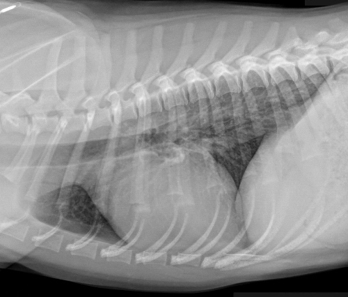
#XRAY BRONCHITIS SERIES#
With this technique there are gaps in the viewed images therefore a second series should also be provided from the volumetric dataset containing thicker (5mm) but contiguous slices. It is recommended that thin slice images be reconstructed between 1-2 mm thick every 10mm, i.e. Images can then be reconstructed in multiple planes and slice thicknesses. This can be obtained in a few seconds or less thereby minimising respiratory and cardiac motion artefact. Multidetector CT scanners enable volumetric data acquisition with scans obtained through the entire thorax in a single breath-hold. Lobar atelectasis (secondary to mucous plugging) Fig 1Ĭompensatory overinflation of the less affected lobe/lung Fig 1 Ring opacities (dilated end-on bronchi) Fig 1 Tubular opacities (mucous filled bronchi) Fig 2 Parallel line opacities (tram tracking) Fig 1a, b Implies non uniform bronchial dilatation. The dilated airway lies adjacent to a pulmonary artery branch giving the appearance of a ring

This describes airways viewed in a transverse plane. This description applies to dilated airways seen in a horizontal orientation. These only describe the appearance of the involved airways but do not elude to a specific cause. Its role has largely been reduced to surveillance for intercurrent infection, progressive lobar collapse or suspected development of cavitary disease in patients with known bronchiectasis.Ĭertain descriptive terms have been used in reporting of bronchiectasis. The CXR in affected individuals is often normal or shows non specific findings. In addition to making the diagnosis, the pattern of disease on HRCT may enable one to limit the differential to a single/few specific causative entities. HRCT is the most sensitive and specific non-invasive method for diagnosing bronchiectasis. High-resolution computed tomography (HRCT) is the cornerstone in the radiological diagnosis of clinically suspicious cases. Imaging plays a pivotal role in the diagnosis of bronchiectasis. Bronchiectasis is defined as irreversible dilatation of a portion of the bronchial tree. The three most important mechanisms that contribute to the pathogenesis of bronchiectasis are infection, airway obstruction and peribronchial fibrosis.


 0 kommentar(er)
